


Abstract
The heart is a nonlinear biological system that can exhibit complex electrical dynamics, complete with period-doubling bifurcations and spiral and scroll waves that can lead to fibrillatory states that compromise the heart's ability to contract and pump blood efficiently. Despite the importance of understanding the range of cardiac dynamics, studying how spiral and scroll waves can initiate, evolve, and be terminated is challenging because of the complicated electrophysiology and anatomy of the heart. Nevertheless, over the last two decades advances in experimental techniques have improved access to experimental data and have made it possible to visualize the electrical state of the heart in more detail than ever before. During the same time, progress in mathematical modeling and computational techniques has facilitated using simulations as a tool for investigating cardiac dynamics. In this paper, we present data from experimental and simulated cardiac tissue and discuss visualization techniques that facilitate understanding of the behavior of electrical spiral and scroll waves in the context of the heart. The paper contains many interactive media, including movies and interactive two- and three-dimensional Java appletsDisclaimer: IOP Publishing was not involved in the programming of this software and does not accept any responsibility for it. You download and run the software at your own risk. If you experience any problems with the software, please contact the author directly. To the fullest extent permitted by law, IOP Publishing Ltd accepts no responsibility for any loss, damage and/or other adverse effect on your computer system caused by your downloading and running this software. IOP Publishing Ltd accepts no responsibility for consequential loss..
Export citation and abstract BibTeX RIS
Figure 2. (Java applet) Human torso with heart in VF
Figure 6(a) and (b). (Java applets) Examples of ionic models of cardiac electrophysiology.
Figure 6(a) and (b). (Java applets) Examples of ionic models of cardiac electrophysiology.
Figure 7. (Java applet) Interactive structure of the heart.
Figure 8(a)–(d). (Java applets) Images of 3D cardiac structures.
Figure 8(a)–(d). (Java applets) Images of 3D cardiac structures.
Figure 8(a)–(d). (Java applets) Images of 3D cardiac structures.
Figure 8(a)–(d). (Java applets) Images of 3D cardiac structures.
Figure 10. (Java applet) Blood vessels in the heart.
Figure 12. (0.7 MB, GIF) Experimental (left) and simulated (right) anatomical reentrant arrhythmia.
Figure 12(a). (4.5 MB, MOV) Experimental anatomical reentrant arrhythmia.
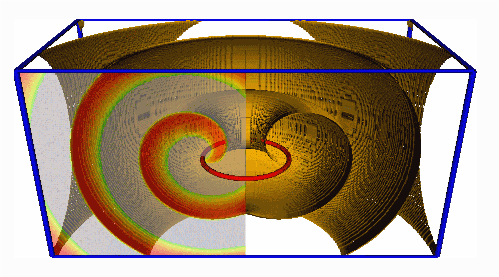
Figure 13. (Java applet) Example of reentry in a 1D ring.
Figure 19. (Java applets) Maps of alternans in (a)–(b) voltage, and (c) calcium.
Figure 19. (Java applets) Maps of alternans in (a)–(b) voltage, and (c) calcium.
Figure 19. (Java applets) Maps of alternans in (a)–(b) voltage, and (c) calcium.
Figure 20. (Java applet) Spatially discordant alternans in a simulated 1D cable.
Figure 21. (2.8 MB, MOV) Spatially discordant alternans progressing to spiral wave breakup.
Figure 22. (2.5 MB, GIF) 'Mother rotor' with fibrillatory conduction and breakup.
Figure 23. (8.8 MB, GIF) Breakup of reentrant waves in canine left ventricle.
Figure 27. (1.2 and 2.0 MB, GIF) Spiral wave dynamics of the ten Tusscher et al model.
Figure 27. (1.2 and 2.0 MB, GIF) Spiral wave dynamics of the ten Tusscher et al model.
Figure 29. (Java applets) Breakup of scroll waves in a simulated 3D tissue slab.
Figure 29. (Java applets) Breakup of scroll waves in a simulated 3D tissue slab.
![Figure 32. (1.4 and 1.1 MB, GIF) (a) Single spiral wave in the human atrial geometry of [76] simulating AFl. (b) Multiple spiral waves in the same geometry simulating AF.](https://content.cld.iop.org/journals/1367-2630/10/12/125016/revision1/Figure_32a_movie.gif)
![Figure 32. (1.4 and 1.1 MB, GIF) (a) Single spiral wave in the human atrial geometry of [76] simulating AFl. (b) Multiple spiral waves in the same geometry simulating AF.](https://content.cld.iop.org/journals/1367-2630/10/12/125016/revision1/Figure_32b_movie.gif)
Figure 33. (Java applets) Arrhythmias in the context of the whole heart.
Figure 33. (Java applets) Arrhythmias in the context of the whole heart.
Figure 33. (Java applets) Arrhythmias in the context of the whole heart.
Figure 33. (Java applets) Arrhythmias in the context of the whole heart.
Figure 34. (1.1 MB, MOV) Termination of equine AF by quinidine.
Figure 38. (1.5 MB, GIF) Visualization of ventricular arrhythmias.